Please see Cell Cycle and Cell Division Class 11 Biology Revision Notes provided below. These revision notes have been prepared as per the latest syllabus and books for Class 11 Biology issues by CBSE, NCERT, and KVS. Students should revise these notes for Chapter 10 Cell Cycle and Cell Division daily and also prior to examinations for understanding all topics and to get better marks in exams. We have provided Class 11 Biology Notes for all chapters on our website.
Chapter 10 Cell Cycle and Cell Division Class 11 Biology Revision Notes
Introduction :
Growth and reproduction are characteristics of cells, indeed of all living organisms. All cells reproduce by dividing into two, with each parental cell giving rise to two daughter cells each time they divide. These newly formed daughter cells can themselves grow and divide, giving rise to a new cell population that is formed by the growth and division of a single parental cell and its progeny. In other words, such cycles of growth and division allow a single cell to form a structure consisting of millions of cells.
MITOSIS
Term mitosis was proposed by Flemming. Mitosis produced genetically identical cells, which are similar to mother cell.
Cause of mitosis :
(I) Kern plasm theory : Hertwig proposed kern plasm theory. According to this theory mitosis occurs due to disturbance in Karyoplasmic Index (KI) or Nucleocytoplasmic ratio of cell.
Karyoplasmic Index : KI = Vn / Vc – Vn
Vn= Volume of nucleus
Vc = Volume of cell
Vc Vn= Volume of cytoplasm
♦ Karyoplasmic Index of small cell is high as they have less cytoplasm. Nucleus efficiently controls the acitivity of cytoplasm in small cells.
♦ In a large cell nucleus fail to control the activity of cytoplasm. To attain the control of nucleus on metabolism a large cell divides into two cells.
(II) Surface-volume Ratio : Surface-volume ratio is also considered as a cause of cell division. When a cell grows in size its volumes increases more than its surface. So a stage will reach when the surface area becomes insufficient to draw the material. At such critical stage, division of cell started.
CELL CYCLE
♦ Cell division is a very important process in all living organisms. During the division of a cell, DNA replication and cell growth also take place.
♦ All these processes, i.e., cell division, DNA replication, and cell growth, hence, have to take place in a coordinated way to ensure correct division and formation of progeny cells containing intact genomes.
♦ The sequence of events by which a cell duplicates its genome, synthesises the other constituents of the cell and eventually divides into two daughter cells is termed cell cycle.
♦ Although cell growth (in terms of cytoplasmic increase) is a continuous process, DNA synthesis occurs only during one specific stage in the cell cycle.
♦ The replicated chromosomes (DNA) are then distributed to daughter nuclei by a complex series of events during
cell division. These events are themselves under genetic control.
PHASES OF CELL CYCLE
♦ A typical eukaryotic cell cycle is illustrated by human cells in culture. These cells divide once in ap- proximately every 24 hours.
♦ Yeast can progress through the cell cycle in only about 90 minutes. The time period of cell cycle is varied from organism to organism and also from cell type to cell type.
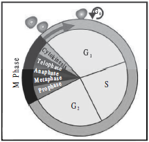
Cell cycle involves two stages :-
1. Interphase
2. Division phase/M-phase
1. Interphase :
This is phase between two successive M-phase. In interphase cell grows in size and prepares itself for next division. Interphase is most active phase of cell cycle. The interphase last more than 95% of the duration of cell cycle.
♦ A series of metabolic changes occurs during interphase in cell. These changes were not visible under micro- scope, So some scientist termed interphase as resting phase. It is the time during which cell is preparing for division by undergoing both cell growth and DNA replication in an orderly manner.
♦ Howard and Pelc classified interphase into three sub stages :-
(i) G1 phase or Pre DNA synthesis phase (Ist Gap phase)
♦ G1 phase corresponds to the interval between mitosis and initiation of DNA replication. During G1 phase the cell is metabolically active and continuously grows
♦ During G1 -most of cell organelles increases in cell and cell rapidly synthesizes different types of RNA and proteins. Due to availability of protein, synthesis of new protoplasm takes place in cell and it starts growing in size. Cell grows maximum in G1 stage.
(ii) S phase (DNA synthesis phase) :
♦ Replication of nuclear DNA and synthesis of histone protein takes place in s-phase. Replication of cytoplasmic DNA may occur in any stage of cell cycle.
♦ During this time the amount of DNA per cell doubles. If the initial amount of DNA is denoted as 2C then it increases to 4C. However, there is no increase in the chromosome number; if the cell had diploid or 2n number of chromosomes at G1 , even after S phase the number of chromosomes remains the same, i.e., 2n.
♦ S-phase marks the phase of DNA replication and chromosome duplication (DNA content in a chromosome become double).
♦ In animal cells, during the S phase, DNA replication begins in the nucleus, and the centriole duplicates in the cytoplasm.
(iii) G2 phase (2nd Gap phase) or Post DNA synthesis phase (Pre mitosis phase)
♦ Actual preparation (Final preparation) of M-phase occurs during this phase. Special materials required for M-phase are synthesized in G2 phase. eg. Tubulin protein. (Required for formation of
spindle fibres). Cell growth continues.
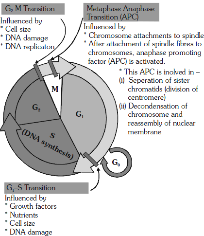
G0 phase –
♦ Some cells in the adult animals do not appear to exhibit division (e.g., heart cells) and many other cells divide only occasionally, as needed to replace cells that have been lost because of injury or cell death. These cells that do not divide further exit G1 phase to enter an inactive stage called quiescent stage (G0) of the cell cycle.
♦ Cells in this stage remain metabolically active but no longer proliferate (divide) unless called on to do so depending on the requirement of the organism.
Checkpoints of cell cycle :
♦ Cell cycle is running by a group of special proteins “Cyclins and Cdks (MPF). (Nurse, T.Hunt & Hartwell 2001 studies on saccharomyces)
♦ Cell cycle is running by a group of special proteins “Cyclins and Cdks.
♦ The activity of enzymes, known as cyclin dependant kinases. (Cdk’s) regulates the cell cycle. Kinase is an enzyme that removes a phosphate group from ATP & add to another protein. The kinases involved in the cell cycle are called Cdks because they are activated when they combined with key protein called cyclin.
♦ At some check points (G1 → S / G2 → M ) a kinase enzyme combines with cyclin & this moves the cell cycle forwardly.
♦ G2M transition is triggered by maturation promoting factor (MPF) formed by M-cyclin + CDK
2. Division phase :
♦ Division phase or Mphase or mitotic phase lasts for only about an hour in the 24 hour duration of cell cycle
of a human cell.
♦ The M-phase represents the phase when the actual cell division or mitosis occurs.
♦ In animals, mitotic cell division is restricted or only seen in diploid somatic cell except in some social insects. Against this, the plants can show mitotic division in both haploid and diploid cells.
♦ This is the most dramatic period of the cell cycle, involving a major reorganisation of virtually all components of the cell. Since the number of chromosomes in the parent and progeny cells is the same, it is also called as equational division.
♦ Though for convenience mitosis has been divided into four stages of nuclear division, it is very essential to understand that cell division is a progressive process and very clear-cut lines cannot be drawn between various stages.
♦ The M-phae start with nuclear division, corresponding to the separation of daughter chromosome (Karyokinesis) and usually ends with division of cytoplasm (cytokinesis).
Mitosis is divided into the following four stages :-
1. Prophase
2. Metaphase
3. Anaphase
4. Telophase
1. Prophase :
♦ Prophase which is the first stage of karyokinesis of mitosis follows the S and G2 phases of interphase.
♦ In the S and G2 phases the new DNA molecules formed are not distinct but interwined.
♦ Prophase is marked by the initiation of condensation of chromosomal material. The chromosomal material becomes untangled during the process of chromatin condensation.
♦ The centriole, which had undergone duplication during S phase of interphase, now begins to move towards opposite poles of the cell.
♦ Formation of astral ray occurs due to gelation of proteins around centrioles in animal cells.
♦ Anastral and Amphiastral Mitosis : In higher plants, centrioles are absent and no asters are formed. Mitosis without asters is known as anastral mitosis. In animals, the asters are present and the mitosis is described as amphiastral or astral mitosis.
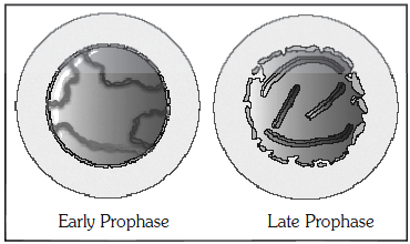
♦ The completion of prophase can thus be marked by the following characteristic events:
a. Chromosomal material condenses to form compact mitotic chromosomes. Chromosomes are seen to be composed of two chromatids attached together at the centromere.
b. Centrosome which had undergone duplication during interphase, begins to move towards opposite poles of the cell. Each centrosome radiates out microtubules called asters. The two asters together with spindle fibres forms mitotic apparatus.
c. Cell at the end of prophase when viewed under the microscope, do not show golgi complexes, endoplasmic reticulum, nucleolus and nuclear envolope.
2. Metaphase :
♦ The complete disintegration of the nuclear envelope marks the start of the second phase of mitosis, hence the chromosomes are spread through the cytoplasm of the cell.
♦ By this stage, condensation of chromosomes is completed and they can be observed clearly under the microscope. This then, is the stage at which morphology of chromosomes is most easily studied.
♦ At this stage, metaphase chromosome is made up of two sister chromatids , which are held together by the centromere . small disc – shaped structures at the surface of the entromeres are called kinetochores. These structures serve as the sites of attachment of spindle fibres (formed by the microtubules) to the chromosomes that are moved into position at the centre of the cell.

♦ Hence, the metaphase is characterised by all the chromosomes coming to lie at the equator with one chromatid of each chromosome connected by its kinetochore to spindle fibres from one pole and its sister chromatid connected by its kinetochore to spindle fibres from the opposite pole. The plane of alignment of the chromosomes at metaphase is referred to as the metaphase plate.
♦ Chromosomal fibres (discontinous/kinetochore which run from pole to centromere) and supporting fibres (continous /non-kinetochore, which run from pole to pole) arrange in cell.
♦ Centromere lies at equator and arms of chromosomes remain directed towards poles.
♦ The key features of metaphase are:
a. Spindle fibres attach to kinetochores of chromosomes.
b. Chromosomes are moved to spindle equator and get aligned along metaphase plate through spindle fibres to both poles.
3. Anaphase :
♦ Centromere of each chromosome splits simultaneously lengthwise (division of centromere). Sister chromatids separate from each other and separated each chromatid is now reffered to as individual chromosome.
♦ Number of chromosome become double in cell.
♦ As each chromosome moves away from the equatorial plate, the centromere of each chromosome is towards the pole and hence at the leading edge, with the arms of the chromosome trailing behind.
♦ The two new daughter chromosomes begin moving toward opposite ends of the cell as their kinetochore microtubule shorten due to depolymerisation of tubulin protein towards kinetochoric end. Because these mircotubules are attached at the centromere region, the centromeres are pulled ahead of the arms. (Pulling)
♦ The cell elongates as the nonkinetochore microtubules lengthen.
♦ Anaphase stage is characterised by the following key events:
a. Centromeres split and chromatids separate.
b. Chromatids (now reffered as chromosomes) move to opposite poles.

4. Telophase (Reverse prophase) :
At the beginning of the final stage of karyokinesis, i.e., telophase, the chromosomes that have reached their respective poles decondense and lose their individuality. The individual chromosomes can no longer be seen and chromatin material tends to collect at each of the two poles. This is the stage which shows the following key events:
a. Chromosomes cluster at opposite spindle poles and their identity is lost as discrete elements.
b. Nuclear envelope develops around the chromosome clusters at each pole forming two daughter nuclei.
c. Nucleolus, golgi complex and ER reform.
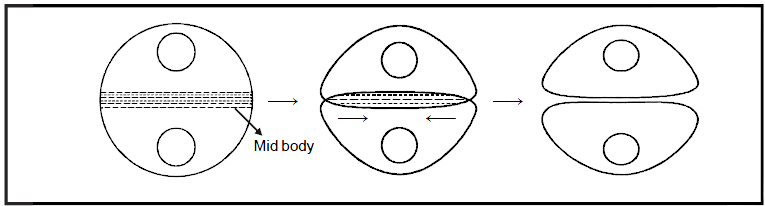
CYTOKINESIS
♦ Mitosis accomplishes not only the segregation of duplicated chromosome into daughter nuclei (Karyokinesis) but the cell itself is divided into two daughter cells by the separation of cytoplasm called cytokinesis at the end of which cell division gets completed
♦ In animals cytokinesis occurs by constriction & furrow formation. Microtubules and microfilaments arrange on equator to form midbody and at the periphary of the equator a contractile ring is formed that is made up of actin and myosin protein. Due to interaction between actin and myosin ring contract, thus a furrow forms from outside to inside in cell. Furrow deepens continuosly and ultimately a cell divides into two daughter cells. In animals cytokinesis occurs in centripetal order.
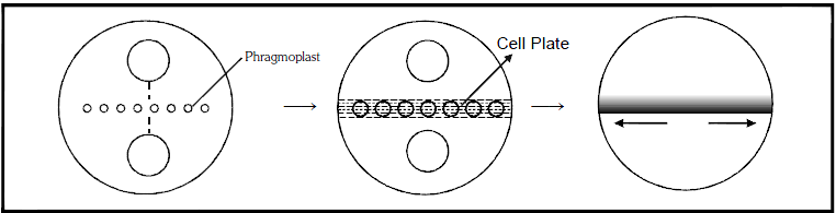
♦ Cytokinesis in plants takes place by cell plate formation because constriction is not possible due to presence
of the rigid cell wall. Many golgi vesicles and spindle microtubules arrange themselves on equator to form phragmoplast. Fragementes of ER may also deposit in phragmoplast. Membrane of golgi vesicles fuse to form a plate like structure called cell plate. Golgi vesicles secret calcium and magnesium pectate. Further cell plate is modified into middle lamella. In plants, cytokinesis occurs in centrifugal order (cell plate formation is from center to periphery).
♦ In some organisms karyokinesis is not followed by cytokinesis as a result of which multinucleate condition arises leading to the formation of syncytium (e.g., liquid endosperm in coconut).
SIGNIFICANCE OF MITOSIS
1. Development of an organism occurs by mitosis. Every organism starts its life from a single cell i.e. zygote. Repeated mitosis in zygote leads to the formation of the whole body.
2. The growth of multicellular organisms is due to mitosis.
3. Cell growth results in disturbing the ratio between the nucleus and the cytoplasm. It therefore becomes essential for the cell to divide to restore the nucleo-cytoplasmic ratio.
4. A very significant contribution of mitosis is cell repair. The cells of the upper layer of the epidermis, cells of the lining of the gut, and blood cells are being constantly replaced.
5. Mitotic divisions in the meristematic tissues the apical and the lateral cambium, result in a continuous growth of plants throughout their life.
MODIFICATIONS OF MITOSIS
1. Free nuclear division : Karyokinesis is not followed by cytokinesis as a result of which multinucleated condition arises .
2. Endomitosis : This is duplication of chromosomes without division of nucleus. Endomitosis leads to polyploidy. i.e. Increase in number of set of chromosomes. Colchicine induces polyploidy in plants. Colchicine is a mitotic poison as it arrests the formation of spindle fibres.
3. Endoreduplication : Endoreduplication is a modification of endomitosis. The polytene chromosomes are formed by the process of endoreduplication. In endoreduplication, the chromatids replicate but do not get seperated. This process is also known as polyteny.
GOLDEN KEY POINTS
♦ During the division of a cell, DNA replication and cell growth take place.
♦ The sequnce of events by which a cell duplicates its genome, synthesis the other constituents of the cell and eventually divides into two doughter cells is termed cell cycle.
♦ The interphase lasts more than 95% of the duration of cell cycle.
♦ S phape marks the phase of DNA replication and chromosome duplication.
♦ In prophase chromosomal material (Chromatin) condenses to form compact mitotic chromosome.
♦ In metaphase spindle fiberes attach to kinetochores of chromosome.
♦ In anaphase centromeres split and chromatids separate.
♦ In animal cell cytokinesis occurs by furrow formation and in plant cell occurs by cell plate method.
♦ A very significant contribution of mitosis is cell repair.
MEIOSIS
♦ “Term meiosis” was proposed by Farmer and Moore.
♦ The specialised kind of cell division that reduces the chromosome number by half results in the production of haploid daughter cells. This kind of division is called meiosis.
♦ Meiosis ensures the production of haploid phase in the life cycle of sexually reproducing organisms whereas fertilisation restores the diploid phase. Meiosis occurs during gametogenesis, leads to the formation of haploid gametes.
The key features of meiosis are as follows:
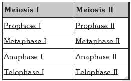
Meiosis involves two sequential cycles of nuclear and cell division called meiosis I and meiosis II but only a single cycle of DNA replication.
Meiosis I :
♦ Heterotypic division or reduction division. It leads to reduction in chromosome numbers. Division of
chromosome does not occurs in meiosis-I and only segregation of homologous chromosomes takes place.
♦ Meiosis I is initiated after the parental chromatids have replicated to produce identical sister chromatids at the S phase.
♦ Meiosis I involves pairing of homologous chromosomes and recombination between non sister chromatids of homologous chromosome.
Meiosis II :
♦ This is a homotypic division or equational division. It does not leads to any change in chromosome number.
♦Division of chromosome or centromere occurs during meiosis II.
♦Four haploid cells are formed at the end of meiosis II. All the four daugther cells produced by meiosis
are genetically diffrent from each other and also differ from the mother cell.
In meiosis, division of nucleus takes place twice but division of chromosome occurs only once in meiosis-II.
Meiotic events can be grouped under the following phases :
Interphase : same as in mitosis
Stages of meiosis I
1. Prophase I :
♦ Typically longer and more complex when compared to prophase of mitosis. Prophase I is classified in five substages based on chromosomal behaviour :
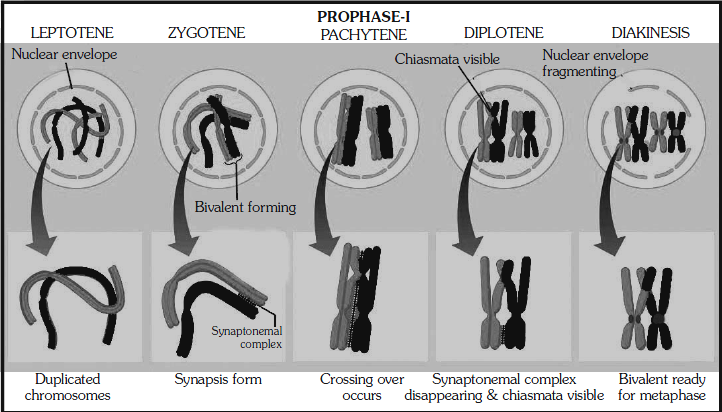
(a) Leptotene: Chromatin threads condense to form chromosomes. Chromosomes are longest & thinest. Chromosomes become gradually visible under the light microscope.
♦ All the chromosomes in nucleus remain directed towards centrioles, so group of chromosomes in nucleus appears like a bouquet. (Bouquet stage)
(b) Zygotene or Synaptotene: Zygotene is characterized by pairing of homologous chromosomes (Synapsis). Pairs of homologous chromosomes are called Bivalents or tetrads. However these are more clearly visible at next stage (pachytene) A structure develops in between homologous chromosomes, Which is termed as synaptonemal complex.
♦ The 1st two stages of prophase I is relatively short lived compared to the pachytene.
(c) Pachytene (Thick thread): Due to increased attraction, homologous chromosomes tightly coil around each other. Both the chromatids of each chromosome become distinct and are called sister chromatids.
♦ During this stage, the four chromotids of each bivalent chromosome become distinct and clearly appeared as tetrad.
♦ Recombination nodules between nonsister chromatids of homologous pair develop and these non sister chromatid exchange their parts i.e. crossing over.
♦ Crossing over leads to recombination of genetic material on the two chromosomes.
♦ Crossing over is an enzyme mediated process and the enzyme involved is called recombinase (Endonuclease + ligase)
♦ Recombination between homologous chromosomes is completed by the end of pachytene, leaving the
chromosomes linked at the sites of crossing over.
(d) Diplotene : The begining of dipotene is recognised by dissolution of synaptonemal complex. Homologous chromosomes start repulsing each other so X-shape structures appeard called chiasmata.
♦ Diplotene may last long up to months or years in oocytes of some vertebrates (Dictyotene).
(e) Diakinesis : It is final stage of meiotic prophase I. Marked by terminalization of chiasmata (Chiasmata open in zip like manner).
♦ Chromosome are fully condensed and meiotic spindle is assembled to prepare the homologous chromosome for separation.
♦ Centrioles move towards the opposite poles.
♦ By the end of diakinesis nucleolus disappear and the nuclear envelop also breaks down.
♦ Diakinesis represents transition to metaphase.
2. Metaphase I :
♦ Bivalents arrange on equator (congression) of cell to form metaphase plate. The microtubules (spindle fibres) from the opposite poles of the spindle attach to the pair of homologous chromosome with one kinetochore of each chromosome.
♦ Two types of spindle fibres appear in the cell :-
(i) Chromosomal / Kinetochore Spindle fibres
(ii) Supporting / Continuous / non-kinetochore Spindle fibres
3. Anaphase I :
♦ Due to shortening of kinetochore/chromosomal fibres homologous chromosomes segregate from each other and move towards the opposite poles. Sister chromatids remain associated at their centromeres (i.e. chromo- somes remain in double chromatid stage)
♦ Anaphase I is characterised by segregation or disjunction of chromosomes. Division of centromere is
absent.
4. Telophase I :
♦ The nuclear membrane and nucleolus reappear. Although in many case the chromosomes do undergo some dispersion, but they do not reach the extremely extended state of the interphase nucleus.
♦ Cytokinesis follows telophase-I and a diploid (2n) cell divides into two haploid (n) daugther cells. This is called as dyad of cells.

♦ Interkinesis : Gap between meiosis I and meiosis II is called Interkinesis. Preparations of meiosis II occur during interkinesis. It is like interphase of mitosis but replication of DNA is absent in interkinesis.
♦ Interkinesis is generally short lived. Interkinesis is followed by prophase-II, a much simpler prophase than
prophase-I.
Stages of Meiosis II
1. Prophase II:
Meiosis II is initiated immediately after cytokinesis, usually before the chromosomes have fully elongated. In contrast to meiosis I, meiosis II resembles a normal mitosis. The nuclear membrane disappears by the end of prophase II. The chromosomes again become compact.
2. Metaphase II:
At this stage the chromosomes align at the equator and the microtubules from opposite poles of the spindle get
attached to the kinetochores of sister chromatids.
3. Anaphase II:
It begins with the simultaneous splitting of the centromere of each chromosome (which was holding the sister chromatids together), allowing them to move toward opposite poles of the cell by shortening of microtubules attached to kinetochores.
4. Telophase II:
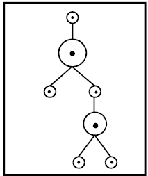
Meiosis ends with telophase II, in which the two groups of chromosomes once again get enclosed by a nuclear envelope; cytokinesis follows resulting in the formation of tetrad of cells i.e., four haploid daughter cells.

Significance of Meiosis :
1. Meiosis is the mechanism by which conservation of specific chromosome number of each species is
chieved across generations in sexually reproducing organisms, even though the process (per se
paradoxically) results in reduction of chromosome number by half.
2. It also increases the genetic variability in the population of organisms from one generation to the
next. Variations are very important for the process of evolution.
GOLDEN KEY POINTS
♦ Porophase-I furthure subdivide into five phases based on the chromosomes behaviour.
♦ Meiosis ensures the production of haploid phase in the life cycle of sexually reproducing organism.
♦ Meiosis involves pairing of homologous chromosomes and recombination betwen them.
♦ Chiasmata formation is the result of crossing over.
♦ Meiosis increases the genetic variability in the population of organism from one generation to next.
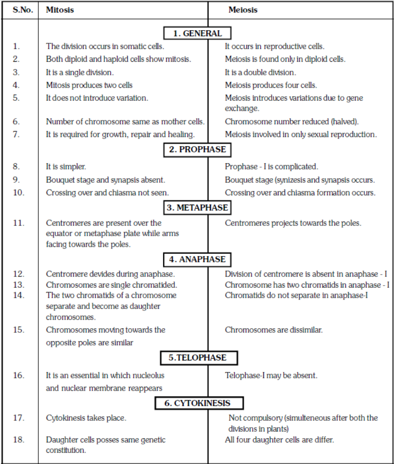
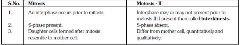
DIFFERENCES BETWEEN MITOSIS AND MEIOSIS