Please see Breathing and Exchange of Gases Class 11 Biology Revision Notes provided below. These revision notes have been prepared as per the latest syllabus and books for Class 11 Biology issues by CBSE, NCERT, and KVS. Students should revise these notes for Chapter 17 Breathing and Exchange of Gases daily and also prior to examinations for understanding all topics and to get better marks in exams. We have provided Class 11 Biology Notes for all chapters on our website.
Chapter 17 Breathing and Exchange of Gases Class 11 Biology Revision Notes
Respiration: Respiration is the oxidation of nutrients in the living cells to release energy for biological work.
Breathing: Breathing is the exchange of O2 from the atmosphere with CO2 produced by the cells.
RESPIRATORY ORGANS
♦ General body surface: E.g. lower invertebrates (sponges, coelenterates, flatworms etc).
♦ Skin or moist cuticle (cutaneous respiration): E.g. earthworms, leech, amphibians etc.
♦ Tracheal tubes: E.g. insects, centipede, millipede, spider.
♦ Gills (Branchial respiration): E.g. fishes, tadpoles, prawn.
♦ Lungs (Pulmonary respiration): E.g. most vertebrates
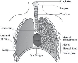
HUMAN RESPIRATORY SYSTEM
It consists of a pair of air passages (air tract) and lungs.
1. Air passages
♦ Conducting part which transports the atmospheric air into the alveoli, clears it from foreign particles, humidifies and brings the air to body temperature.
External nostrils → nasal passage → nasal chamber
(cavity) → pharynx → glottis → larynx → trachea →
primary bronchi → secondary bronchi → tertiary bronchi
→ bronchioles → terminal bronchioles → respiratory
bronchiole → alveolar duct.
♦ Each terminal bronchiole gives rise to many very thin and vascularised alveoli (in lungs).
♦ A cartilaginous Larynx (sound box or voice box) helps in sound production.
♦ During swallowing, epiglottis (a thin elastic cartilaginous flap) closes glottis to prevent entry of food into larynx.
♦ Trachea, all bronchi and initial bronchioles are supported by incomplete cartilaginous half rings.
2. Lungs
♦ Lungs situate in thoracic chamber and rest on diaphragm.
♦ Right lung has 3 lobes and left lung has 2 lobes.
♦ Lungs are covered by double-layered pleura (outer parietal pleura and inner visceral pleura).
♦ The pleural fluid present in between these 2 layers lubricates the surface of the lungs and prevents friction between the membranes.
♦ Lungs= Bronchi + bronchioles + alveoli.
♦ Alveoli and their ducts form the respiratory or exchange part of the respiratory system.
♦ Alveoli are the structural and functional units of lungs.
Steps of respiration
1. Pulmonary ventilation (breathing).
2. Gas exchange between lung alveoli & blood.
3. Gas transport (O2 transport & CO2 transport).
4. Gas exchange between blood & tissues.
5. Cellular or tissue respiration.
MECHANISM OF BREATHING (INSPIRATION & EXPIRATION)
a. Inspiration
♦ Active intake of air from atmosphere into lungs.
♦ During this, the diaphragm contracts (flattens) causing an increase in vertical thoracic volume (antero-posterior axis).
♦ Contraction of external intercostal muscles (muscles found between ribs) lifts up the ribs and sternum causing an increase in thoracic volume in the dorso-ventral axis.
♦ Increase in thoracic volume reduces thoracic pressure. So, lungs expand. Thus, pulmonary volume increases resulting in decrease of intra-pulmonary pressure to less than the atmospheric pressure. So, air moves into lungs.

b. Expiration
♦ Passive expelling of air from the lungs.
♦ During this, intercostal muscles & diaphragm relax causing a decrease in thoracic volume and thereby pulmonary volume. So, air moves out.
♦ During forceful expiration, abdominal muscles and internal inter-costal muscles contract.
Respiratory volumes and capacities:
Tidal volume (TV): Volume of air inspired or expired during a normal respiration. It is about 500 ml. i.e., 6000- 8000 ml per minute.
Inspiratory reserve volume (IRV) or complemental air: Additional volume of air that can inspire by forceful inspiration. It is 2500-3000 ml.
Expiratory reserve volume (ERV) or supplemental air: Additional volume of air that can expire by a forceful expiration. It is 1000-1100 ml.
Residual volume (RV): Volume of air remaining in lungs after a forcible expiration. It is 1100-1200 ml
Inspiratory capacity (IC): Total volume of air inspired after a normal expiration (TV + IRV). It is 3000-3500 ml.
Expiratory capacity (EC): Total volume of air expired after a normal inspiration (TV + ERV). It is 1500-1600 ml.
Functional residual capacity (FRC): Volume of air remaining in the lungs after a normal expiration (ERV + RV). It is 2100-2300 ml.
Vital capacity (VC): Volume of air that can breathe in after a forced expiration or Volume of air that can breathe out after a forced inspiration (ERV + TV + IRV). It is 3500-4500 ml.
Total lung capacity (TLC): Total volume of air in the lungs after a maximum inspiration. (RV + ERV + TV + IRV or VC + RV). It is 5000-6000 ml.
dead space: Part of respiratory tract (from nostrils to terminal bronchi) not involved in gaseous exchange is called dead space. Dead air volume is about 150 ml.
♦ Respiratory cycle= an inspiration + an expiration
♦ Normal respiratory (breathing) rate: 12-16 times/min
♦ Spirometer (respirometer): To measure respiratory rate
GAS EXCHANGE
Gas exchange occurs between
1. Alveoli and blood
2. Blood and tissues
Alveoli are the primary sites of gas exchange.
O2 & CO2 are exchanged by simple diffusion. It depends upon the following factors:
Pressure/ concentration gradient: The Partial pressures (individual pressure of a gas in a gas mixture) of O2 and CO2 (pO2 and pCO2) are given below.

pO2 in alveoli is more (104 mm Hg) than that in blood capillaries (40 mm Hg). So O2 diffuses into capillary blood. pCO2 in deoxygenated blood is more (45 mm Hg) than that in alveoli (40 mm Hg). So, CO2 diffuses to alveoli.
Solubility of gases: Solubility of CO2 is 20-25 times higher than that of O2. So, the amount of CO2 that can diffuse through the diffusion membrane per unit difference in partial pressure is higher than that of O2.
Thickness of membranes: The diffusion membrane is made up of 3 layers:
a) Squamous epithelium of alveoli.
b) Endothelium of alveolar capillaries.
c) Basement substance between them.
Its total thickness is only 0.5 μm. It enables easy gas exchange.
Surface area: Presence of alveoli increases the surface area of lungs. It increases the gas exchange.

GAS TRANSPORT (O2TRANSPORT & CO2 TRANSPORT)
It is the transport of respiratory gases (O2& CO2) from alveoli to the systemic tissues and vice versa.
1. O2 TRANSPORT
It is the transport of O2 from lungs to various tissues. It occurs in 2 ways:
a. In physical solution (blood plasma): About 3% of O2 is carried in a dissolved state through plasma.
b. As oxyhaemoglobin: About 97% of O2 is transported by haemoglobin (red coloured iron containing pigment) on RBC. O2 binds with haemoglob in (Hb) to form oxyhaemoglobin. This is called oxygenation. Hb has 4 haem units. So, each Hb molecule can carry 4 oxygen molecules. Binding of O2 depends upon pO2 , pCO2 , H+ ion concentration (pH) and temperature.
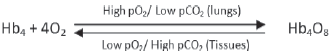
♦ In the alveoli, high pO2 , low pCO2 , lesser H+ ion concentration and lower temperature exist. These factors are favourable for the formation of oxyhaemoglobin.
♦ In tissues, low pO2 , high pCO2 , high H+ ions and high temperature exist. So Hb4O8 dissociates to release O2 .
♦ Every 100 ml of oxygenated blood can deliver around 5 ml of O2 to the tissues under normal physiological conditions.
Oxygen-haemoglobin dissociation curve
It is a sigmoid curve obtained when percentage saturation of Hb with O2 is plotted against the p O2.
It is used to study the effect of factors like pCO2, H+ concentration etc., on binding of O2 with Hb.

2. CO2 TRANSPORT
It is the transport of CO2 from tissues to lungs. In tissues, pCO2 is high due to catabolism and pO2 is low. In lungs, pCO2 is low and pO2 is high. This favours CO2 transport from tissues to lungs. It occurs in 3 ways:
a. As carbonic acid: In tissues, 7% of CO2 is dissolved in plasma water to form carbonic acid and carried to lungs.
b. As carbamino-haemoglobin: In tissues, 20-25% of CO2 binds to Hb to form carbamino-haemoglobin. In alveoli, CO2 dissociates from carbamino-haemoglobin.
c. As bicarbonates: 70% of CO2 transported by this method. RBCs contain an enzyme, carbonic anhydrase. (It is slightly present in plasma too). At tissue site, it facilitates the following reactions:
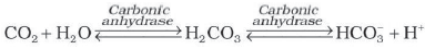
In alveoli, the above reaction proceeds in opposite direction leading to the formation of CO2 and H2O.
Every 100 ml of deoxygenated blood delivers about 4 ml of CO2 to the alveoli.
REGULATION OF RESPIRATION
In brain, there are the following Respiratory centres:
Respiratory rhythm centre (Inspiratory & Expiratory centres): In medulla oblongata. It regulates respiratory rhythms.
Pneumotaxic centre: In Pons. It moderates functions of respiratory rhythm centre. Impulse from this centre reduces the duration of inspiration and thereby alter respiratory rate.
Chemosensitive area: Seen adjacent to the rhythm centre. Increase in the concentration of CO2 and H+ activates this centre, which in turn signals rhythm centre. Receptors in aortic arch & carotid artery also recognize changes in CO2 & H+ concentration and send signals to rhythm centre.Role of oxygen in the regulation of respiratory rhythm is quite insignificant.
DISORDERS OF RESPIRATORY SYSTEM
Asthma: Difficulty in breathing causing wheezing due to inflammation of bronchi and bronchioles.
Emphysema: Damage of alveolar walls. It decreases respiratory surface. Major cause is cigarette smoking.
Occupational respiratory disorders: Certain industries produce so much dust. So, the defense mechanism of the body cannot cope with the situation. Long exposure causes inflammation leading to fibrosis (proliferation of fibrous tissues). It results in lung damage. Workers in such industries should wear protective masks.