Please see Neural Control and Coordination Class 11 Biology Revision Notes provided below. These revision notes have been prepared as per the latest syllabus and books for Class 11 Biology issues by CBSE, NCERT, and KVS. Students should revise these notes for Chapter 21 Neural Control and Coordination daily and also prior to examinations for understanding all topics and to get better marks in exams. We have provided Class 11 Biology Notes for all chapters on our website.
Chapter 21 Neural Control and Coordination Class 11 Biology Revision Notes
♦ Neural (Nervous) system is a system that controls and coordinates the body activities, conducts and integrates the information and responds to stimuli.
♦ It includes brain, spinal cord and nerves.
♦ It is made up of specialized cells known as neurons.
Neuron (nerve cell)
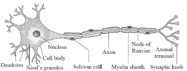
Neuron is the structural and functional unit of neural system. It is composed of
Cell body (cyton): Contains cytoplasm, cell organelles and Nissl’s granules (granular bodies).
Dendron: Short fibres projecting from the cyton. Their sub branches (dendrites) transmit impulses towards the cyton.
Axon: A long fibre which transmit impulses away from the cell body. The branching of axon is called axonite. Each axonite ends as a bulb-like structure called synaptic knob.
Types of Neurons
1. Unipolar: One axon. No Dendron. Found in embryo.
2. Bipolar: One axon and one dendron. Found in the retina.
3. Multipolar: One axon and 2 or more dendrons. Most
common type. Found in the CNS & PNS.
Types of axon
1. Myelinated axon: It is enveloped with Schwann cells that orm a myelin sheath around the axon. Found in spinal & cranial nerves. The white coloured area, formed of myelinated nerve fibres is called white matter. Gaps b/w 2adjacent myelin sheaths are called nodes of Ranvier.
2. Non myelinated axon: Schwann cells present but no myelin sheath. The gray coloured area without myelin sheath is called gray matter. Found in autonomous & somatic neural systems.
GENERATION & CONDUCTION OF NERVE IMPULSES
Impulse transmission is electrochemical. It has 3 steps:
1. Maintenance of resting membrane potential
♦ Neural membrane contains various selectively permeable ion channels.
♦ In a resting neuron (neuron not conducting impulse), the axonal membrane is more permeable to K+ ions and nearly impermeable to Na+ ions. Also, the membrane is impermeable to negatively charged proteins in axoplasm.
♦ Therefore, concentration of K+ and –vely charged proteins in axoplasm is high and concentration of Na+ is low.
♦ The fluid outside the axon contains low concentration of K+ and high concentration of Na+. This forms an ionic or concentration gradient across resting membrane.
♦ The ionic gradients are maintained by the active transport of ions by the Na -K pump. It transports 3 Na+ outwards for 2K+ into the cell. As a result, the outer surface becomes positively charged and inner surface becomes negatively charged (i.e, polarized).
♦ The electrical potential difference across the resting plasma membrane is called as the resting potential.
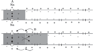
2. Action Potential
♦ When a stimulus is applied, the membrane at the site A becomes permeable to Na+. This causes rapid influx of Na+ and reversal of the polarity at that site (outer negative and inner positive). It is called depolarization.
♦ The electrical potential difference during depolarization across the plasma membrane is called action potential (a nerve impulse).
3. Propagation of action potential
♦ At sites ahead (site B), outer surface is positive and inner surface is negative. As a result, a current flows on the inner surface from site A to site B.
♦ On the outer surface, current flows from site B to site A to complete the circuit. Hence, the polarity is reversed and action potential is generated at site B. i.e., action potential at site A arrives at site B.
♦ The sequence is repeated along the axon and the impulse is conducted.
♦ The rise in permeability to Na+ is extremely short lived. It is quickly followed by a rise in permeability to K+.
♦ Immediately, K+ diffuses outside the membrane and restores the resting membrane. Thus the fibre becomes ready for further stimulation.
Synaptic transmission of impulses
♦ Synapse is a functional junction between two neurons.
♦ It is 2 types: Electrical & Chemical.
1. Electrical synapses
♦ In this, the membranes of pre – and post – synaptic neurons are in close proximity. So impulse transmission is similar to the transmission along an axon.
♦ Impulse transmission is faster than in chemical synapse.
♦ Electrical synapses are very rare in human system.
2. Chemical synapses
♦ In this, there is a fluid filled space (synaptic cleft) between the presynaptic neuron and postsynaptic neuron.
♦ The presynaptic regions have swellings called Synaptic knob (buttons). They contain synaptic vesicles filled with neurotransmitters (acetylcholine or adrenaline).
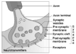
Impulse transmission through chemical synapse:
Impulse reaches at axon terminal → synaptic vesicles bind on plasma membrane → release of neurotransmitter → It diffuses across synaptic cleft → combine with receptors on the post synaptic membrane → opening of ion channels allowing entry of ions → generates action potential.
This action potential may be excitatory or inhibitory.
HUMAN NERVOUS (NEURAL) SYSTEM
It has 2 parts:
Central neural system (CNS): Brain & spinal cord.
Peripheral nervous system (PNS): All nerves.
CENTRAL NEURAL SYSTEM (CNS)
1. BRAIN
♦ It is protected in cranial cavity.
♦ It has 3 – layered connective tissue membranes called cranial meninges.
♦ Meninges consist of outer dura mater, middle arachnoid mater and inner pia mater.
♦ The subarachnoid space (space between pia mater and arachnoid mater) is filled with cerebrospinal fluid (CSF). The ventricles of brain are also filled with CSF.
♦ Brain has 3 divisions: Forebrain, Midbrain & Hindbrain.
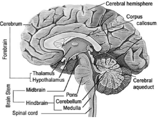
a. Forebrain (Prosencephalon)
It is the anterior part. Consists of cerebrum & diencephalon.
1. Cerebrum
♦ Largest part. It has 2 cerebral hemispheres held together by a tract of nerve fibres (Corpus callosum).
♦ Outer part of cerebrum is called cerebral cortex. It has convulsions & depressions and is formed of gray matter. Gray colour is due to the presence of neuron cell bodies.
♦ Inner part of cerebrum is formed of white matter.
♦ Cerebral cortex consists of
Motor area: Controls voluntary movements of muscles.
Sensory (Somaesthetic) area: Controls the functioning of sense organs.
Association area: It is neither clearly sensory nor motor in function. Responsible for intersensory associations, memory and communication Integrated activities of different centres of cerebral cortex control intelligence, memory, judgment, learning, thinking and articulate speech.
2. Diencephalon (Thalamus & Hypothalamus)
♦ Thalamus: It is the structure around which the cerebrum wraps. It is a coordinating centre (relay station) for sensory and motor impulses.
♦ Hypothalamus: Seen below the thalamus. It
a. Regulates temperature, thirst, hunger and emotions.
b. Secretes hypothalamic hormones.
c. Controls pituitary gland.
d. Controls sleep, wakefulness, blood pressure, heart rate.
♦ The inner parts of cerebral hemispheres and a group of associated deep structures like amygdala, hippocampus, hypothalamus, etc. together constitute Limbic system (Limbic lobe). It regulates sexual behavior, motivations, emotions (excitement, pleasure, rage, fear etc).
b. Midbrain (Mesencephalon)
♦ It is located between thalamus/hypothalamus and Pons.
♦ A canal (cerebral aqueduct) passes through the mid brain.
♦ Mid brain consists of 4 round lobes called Corpora quadrigemina. Their anterior pair is the centre of visual reflexes and the posterior pair is a centre of auditory reflex.
c. Hindbrain (Rhombencephalon)
It consists of cerebellum, Pons & Medulla oblongata. Midbrain & hindbrain form the Brain stem.
♦ Cerebellum (“little cerebrum”): It has very convoluted surface to accommodate more neurons. It co – ordinates muscular activities and body equilibrium.
♦ Pons varoli: It consists of fibre tracts that interconnect different regions of the brain. It co – ordinates the activities of eye and ear and regulates respiration.
♦ Medulla oblongata: It is connected to spinal cord. It controls respiration, cardiovascular reflexes, gastric secretions, peristalsis etc. It also controls salivation,vomiting, sneezing & coughing.
2. SPINAL CORD
♦ It is enclosed within the spinal canal of vertebral column.
♦ It is also protected by meninges.
♦ Spinal cord has a central canal containing CSF.
♦ Outer white matter and inner gray matter.
Functions:
1. Conduction of impulses to and from the brain.
2. Centre of spinal reflexes.
PERIPHERAL NEURAL SYSTEM (PNS)
It includes cranial nerves and spinal nerves.
Nerve fibres of PNS are 2 types:
1. Afferent (sensory) fibres: Carry impulses from sense organs to CNS.
2. Efferent (motor) fibres: Carry impulses from CNS to muscles and glands.
PNS has 2 divisions. They are
1. Somatic neural system: Relays impulses from the CNS to skeletal muscles.
2. Autonomic neural system (ANS): Transmits impulses from CNS to involuntary organs & smooth muscles. It includes sympathetic & parasympathetic nerves. Sympathetic system prepares body to cope with emergencies, stresses & dangers. It increases heartbeat, breathing rate, constricts arteries and elevates BP. Parasympathetic system returns the body to a resting state after stressful situations and slows down heartbeat, dilates arteries, lowers BP etc.
Visceral nervous system is the part of PNS. It includes nerves, fibres, ganglia & plexus by which impulses travel from CNS to the viscera and from viscera to CNS.
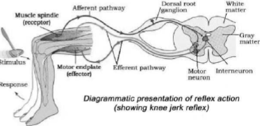
REFLEX ACTION
It is the rapid, involuntary and unconscious actions of body in response to a stimulus. E.g.
♦ Withdrawal of the hand when it touches a hot object.
♦ Touching lips of a nursing baby evokes sucking reflex.
♦ Closing of the eyelids when light falls on them.
♦ Knee jerk phenomenon.
♦ If a child sees or smells a food unknown to him, he does not salivate. But if he sees or smells that food every time before tasting it, he salivates (conditioned reflex).
The pathway of impulses in a reflex action is called Reflex arc. It consists of
1. A receptor organ: It receives the stimulus.
2. Sensory (afferent) neuron: It transmits impulses from sense organ to CNS.
3. Intermediate (connector) neuron: It connects sensory and motor neurons.
4. Motor (efferent/effector/excitor) neuron: It conducts impulse from the CNS to effector organ.
5. An effector organ (muscle/gland): It responds to impulse.
SENSORY RECEPTION & PROCESSING (SENSE ORGANS)
♦ These are the organs that detect the changes in the environment and convey the information to the CNS.
♦ It includes eye, ear, nose, tongue & skin.
EYE
♦ Two eyes are located in sockets of the skull called orbits.
♦ The adult human eyeball is nearly spherical.
♦ Eyeball has three layers: Sclera, Choroid & Retina.
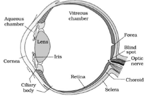
a. Sclera
♦ The external layer formed of a dense connective tissue.
♦ Anterior transparent portion of sclera is called cornea.
b. Choroid
♦ Bluish middle layer. Contains many blood vessels.
♦ Choroid is thin over posterior two – thirds of the eyeball, but it is thick in the anterior part to form ciliary body.
♦ Ciliary body continues forward to form a visible pigmented and opaque portion of the eye called the iris.
♦ Iris has a central opening called pupil. The diameter of the pupil is regulated by the muscle fibres of iris. This helps to regulate the amount of light entering the eye.
♦ The eyeball contains a transparent crystalline lens. It is held in place by ligaments attached to the ciliary body.
c. Retina
♦ Inner layer. It contains 3 layers of cells – from inner to outer – ganglion cells, bipolar cells & photoreceptor cells.
♦ Photoreceptor cells are 2 types: rods and cones. They contain photosensitive proteins (photopigments).
♦ Photopigments are formed of opsin (a protein) and retinal (an aldehyde of vitamin A).
Cone cells:
♦ Function: Daylight (photopic) vision & colour vision.
♦ There are 3 types of cones containing photopigments (photopsin) that respond to red, green and blue lights.
♦ The sensations of different colours are produced by combinations of these cones and their photopigments.
♦ When the cones are stimulated equally, a sensation of white light is produced.
Rod cells:
♦ Function: Twilight (scotopic) vision.
♦ They contain a purplish – red protein called rhodopsin (visual purple). It contains a derivative of Vitamin A.
♦ At the region, slightly above the posterior pole of the eyeball, optic nerves leave the eye and retinal blood vessels enter it. Here, photoreceptor cells are absent. It is called blind spot.
♦ Lateral to the blind spot, there is a yellowish pigmented spot called macula lutea with a central pit (fovea).
♦ The fovea is a thinned – out portion of the retina where only the cones are densely packed. It is the point of greatest visual acuity (resolution).
♦ The space between the cornea and lens is called aqueous chamber. It contains aqueous humor (thin watery fluid).
♦ The space between the lens and retina is called vitreous chamber. It contains vitreous humor (a transparent gel).
Mechanism of vision
Light reflected from an object → enters the eye through cornea & lens → focus on retina → dissociation of retinal from opsin → changes in membrane permeability → generates potential differences (impulse) in photoreceptor cells → generates action potentials in ganglion cells through bipolar cells → impulses are transmitted by optic nerves to brain (visual cortex) → impulses are analyzed and the image is recognized based on memory and experience → vision.
EAR (STATO – ACOUSTIC ORGAN)
♦ It is the organ for hearing & balancing.
♦ It has 3 divisions: External ear, middle ear & inner ear.

External ear
♦ Consists of pinna (ear lobe) & auditory meatus (ear canal).
♦ At the opening of ear canal, hairs are seen.
♦ Ear canal and skin of pinna contains ceruminous glands modified sweat glands). They secrete wax (cerumen).
♦ Wax and hairs protects ears from foreign objects.
♦ Ear canal ends in tympanic membrane (Tympanum or ear drum). It is a semi – transparent membrane covered by a thin layer of skin on itsouter surface and by mucous membrane on the inside.
Middle ear
♦ Consists of tympanic cavity and ear ossicles.
♦ Tympanic cavity is an air filled space that separates the external and inner ear portions.
♦ An auditory tube (Eustachian canal) connects middle ear to the pharynx. It maintains an equal pressure on either side of the eardrum.
♦ Ear ossicles include 3 small bones namely Malleus, Incus and stapes. Malleus is attached to tympanum.
♦ Stapes is the smallest bone of the body. It is attached to membrane of oval window (fenestra ovalis) of inner ear.
Inner ear
♦ It consists of bony labyrinth & membranous labyrinth.
♦ Bony labyrinth is a cavity filled with perilymph.
♦ The membranous labyrinth consists of cochlea and Vestibular apparatus.
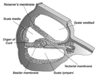
Cochlea (organ of hearing):
♦ It is a coiled structure having 3 canals -upper scala vestibula, middle scala media and lower scala tympani.
♦ Scala vestibula & scala media are separated by Reissner’s membrane.
♦ Scala media and scala tympani are separated by basilar membrane.
♦ S. vestibula & S. tympani are filled with perilymph and scala media is filled with endolymph.
♦ Resting on the basilar membrane and projecting into scala media is complex receptor organ called Organ of Corti. It consists of row of sensory hair cells. The hairs (stereo cilia) of these cells project upwards and lie in contact with tectorial membrane, which projects above them.
Vestibular apparatus:
♦ It consists of 3 semicircular canals and otolith organ.
♦ 2 semicircular canals are vertical and one is horizontal. One end of each canal has a bulging called ampulla. Inside it is a lump called crista ampullaris. Long cilia of cells of crista are grouped together in a bundle (cupula).
♦ Otolith organ consists of utricle and saccule.
♦ Utricle & Saccule have a projecting ridge called macula.
♦ Crista and Macula are specific receptors in vestibular apparatus. They contain sensory hair cells. They are responsible equilibrium & posture of body.
Mechanism of hearing
Pinna collects sound waves → waves reach the tympanic membrane via ear canal → tympanic membrane vibrates → vibrations transmit to ear ossicles & oval window → perilymph in the vestibular canal vibrates → vibrations reach the scala tympani and force the basilar membrane to vibrate → hair endings of sensory hair cells press against tectorial membrane → sensory hair cells are excited → auditory nerve carries impulses to auditory centre of the brain → hearing.
NOSE
♦ Organ of smell (olfaction).
♦ It contains mucus – coated receptors (olfactory receptors) made up of olfactory epithelium. They receive sense of smell. It contains 3 kinds of cells.
♦ The neurons of olfactory epithelium extend from the outside environment directly into a pair of broad beansized organs, called olfactory bulb. These are extensions of the brain’s limbic system.
TONGUE
♦ Organ of taste (gustation).
♦ 4 primary tastes are sweet, salt, sour and bitter.
♦ Taste buds (Gustatoreceptors + supporting cells) are seen around the bases of taste papillae.
Nose & tongue are chemoreceptors (detect dissolved chemicals). Senses of taste & smell are functionally similar and interrelated. The brain integrates different input from taste buds and a complex flavour is perceived
SKIN (Cutaneous receptors)
♦ Largest sense organ.
♦ It contains receptors for heat, cold, touch, pain & pressure.