Please see Body Fluids and Circulation Class 11 Biology Revision Notes provided below. These revision notes have been prepared as per the latest syllabus and books for Class 11 Biology issues by CBSE, NCERT, and KVS. Students should revise these notes for Chapter 18 Body Fluids and Circulation daily and also prior to examinations for understanding all topics and to get better marks in exams. We have provided Class 11 Biology Notes for all chapters on our website.
Chapter 18 Body Fluids and Circulation Class 11 Biology Revision Notes
Circulation is the transport of nutrients, oxygen, CO2 and excretory products to the concerned tissues or organs.
For circulation, simple organisms (sponges, coelenterates etc.) use water from their surroundings. Complex organisms use body fluids (blood & lymph) for circulation.
CIRCULATORY PATHWAYS
Circulatory system is 2 types- Open and Closed.
Open circulatory system: Here, the blood pumped by the heart passes through large vessels into open spaces or cavities called sinuses. E.g. Arthropods and molluscs.
Closed circulatory system: Here, the blood pumped by the heart is always circulated through blood vessels. This is more advantageous as the flow of fluid can be more precisely regulated. E.g. Annelids and chordates.
All vertebrates have a muscular chambered heart.
Fishes: 2-chambered heart (an atrium + a ventricle).
Amphibians: 3-chambered heart (2 atria + a ventricle).
Reptiles (except crocodiles): 3-chambered heart (2 atria + a ventricle). Ventricle is incompletely partitioned.
Crocodiles, birds & mammals: 4-chambered heart.
Types of circulation
Single circulation: In fishes. In this, heart receives impure blood only (venous heart).
Deoxygenated blood → to heart → to gills → oxygenated
blood → to body parts → deoxygenated blood → to heart.
Incomplete double circulation: In amphibians & reptiles. In this, left atrium gets oxygenated blood from gills/ lungs/skin and right atrium gets deoxygenated blood from other body parts. However, they get mixed up in the single ventricle. It pumps out mixed blood.
Double circulation: In birds & mammals. Right atrium gets deoxygenated blood and passes to right ventricle and left atrium gets oxygenated blood and passes to left ventricle. The ventricles pump it out separately without any mixing up.
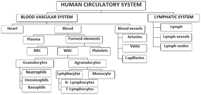
BLOOD VASCULAR SYSTEM
It includes Heart, Blood & Blood vessels.
1. BLOOD
Formed of plasma (55%) & formed elements (45%).
a. PLASMA: Straw-coloured, slightly alkaline (pH 7.4) viscous fluid.
Constituents of plasma:
♦ Water (90-92%): It is a good solvent.
♦ Plasma proteins (6-8 %): Include
Fibrinogen: For blood coagulation.
Globulins: Act as antibodies (for defense of the body).
Albumins: For osmotic balance. Regulate blood pressure.
♦ Glucose, amino acids, lipids & cholesterol.
♦ Inorganic constituents: Na+, Ca2+, Mg2+, Cl–, HCO3– etc.
♦ Gases like CO2, O2, N2 etc.
Plasma without clotting factors is known as Serum.
b. FORMED ELEMENTS (RBC, WBC & PLATELETS)
Red Blood Cells (RBC) or Erythrocytes:
♦ Biconcave non-nucleated cells. No mitochondria, Golgi complex etc. Red colour is due to Haemoglobin (iron containing protein). Normal Hb level is 12-16 g/ 100 ml.
♦ Count: 5 – 5.5 millions/ mm3.
♦ Formed in: Red Bone marrow.
♦ Average lifespan: 120 days. Worn-out RBCs are destroyed in spleen (graveyard of RBCs).
♦ Function: CO2 and O2 transports.
White Blood Cells (WBC) or Leucocytes:
♦ Colourless nucleated cells.
♦ Count: 6000-8000 /mm3.
♦ Formed in: Bone marrow, lymph glands, spleen.
♦ Average lifespan: Generally short lived (1- 15 days).
♦ Function: Part of immune system.
Types of WBC: Granulocytes & Agranulocytes
1. Granulocytes
They are 3 types:
a. Neutrophils (Heterophils): 60-65%. Soldier of the body.
Function: Phagocytosis.
b. Eosinophils (Acidophils): 2-3%. Resist infections. Cause allergic reactions.
c. Basophils (Cyanophils): 0.5-1%. Secrete histamine, serotonin, heparin etc. Cause inflammatory reactions.
2. Agranulocytes
They are 2 types:
a. Lymphocytes (20-25%): Smallest WBC with largest nucleus. Includes B– lymphocytes & T–lymphocytes. Cause immune responses. Secrete antibodies.
b. Monocytes (6-8%): Largest WBC.
Function: Phagocytosis.
Platelets (Thrombocytes):
♦ Colourless non–nucleated cell fragments.
♦ Count: 1.5– 3.5 lakhs /mm3.
♦ Formed in: Megakaryocytes in Bone marrow.
♦ Average lifespan: 7 days.
♦ Function: Blood clotting.

BLOOD COAGULATION
It is a mechanism for haemostasis (prevention of blood loss through injuries). At the site of injury, following events occur:
Clumped platelets & tissues release thromboplastin → It forms thrombokinase (Prothrombinase) enzyme → Thrombokinase hydrolyses prothrombin to thrombin enzyme in presence of Ca2+ → Thrombin converts soluble fibrinogen to insoluble fibrin → Fibrin threads trap dead & damaged blood cells to form clot (coagulum).
BLOOD GROUPS
Blood groups were discovered by Carl Land Steiner.
a. ABO grouping
It is based on presence or absence of 2 surface antigens (chemicals that induce immune response) on RBCs namely A & B. Similarly, plasma contains 2 antibodies (proteins produced in response to antigens) namely anti-A & anti-B.

♦ Antigen A reacts with anti-A. Antigen B reacts with anti-B.
♦ If bloods with interactive antigens & antibodies are mixed together, it causes clumping (agglutination) of RBCs.
♦ Persons with O Group are called Universal donors because they can donate blood to persons with any other blood group. Persons with AB group are called Universal recipients because they can accept blood from all groups.
b. Rh grouping
♦ Rhesus (Rh) factor is another antigen found on RBC.
♦ Rh+ve means the presence of Rh factor and Rh-ve means absence of Rh factor. Nearly 80% of humans are Rh+ve.
♦ Anti-Rh antibodies are not naturally found. So Rh-ve person can receive Rh+ve blood only once but it causes the development of anti-Rh antibodies in his blood. So, a second transfusion of Rh+ve blood causes agglutination. Therefore, Rh-group should be matched before transfusion.
Erythroblastosis foetalis
♦ It is a Rh incompatibility between the Rh-ve blood of a pregnant mother and Rh+ve blood of the foetus.
♦ Rh antigens do not get mixed with maternal blood in first pregnancy because placenta separates the two bloods.
♦ But during first delivery, the maternal blood may be exposed to small amount of foetal blood (Rh+ve). This induces the formation of Rh antibodies in maternal blood.
♦ In case of her subsequent pregnancies, the Rh antibodies from the mother leak into the foetal blood (Rh+ve) and destroy the foetal RBCs. This is fatal to the foetus or cause severe anaemia and jaundice to the baby. This condition is called Erythroblastosis foetalis.
♦ It can be avoided by administering anti-Rh antibodies to the mother immediately after the first delivery.
2. BLOOD VESSELS
Blood vessels are 3 types: Arteries, Veins & Capillaries.
♦ Arteries: They carry blood from heart to other tissues. They contain oxygenated blood (except pulmonary artery) . Their smaller branches are called arterioles. Arteries are 3- middle tunica media (smooth muscles & elastic fibres) and outer tunica externa (fibrous connective tissue).
♦ Veins: They carry blood towards heart. They contain deoxygenated blood (except pulmonary vein). Their smaller branches are called venules. Veins are also 3-layered but tunica media is comparatively thin.
♦ Capillaries: In tissues, arterioles divide into thin walled and single layered vessels. They are called capillaries. They unite into venules.
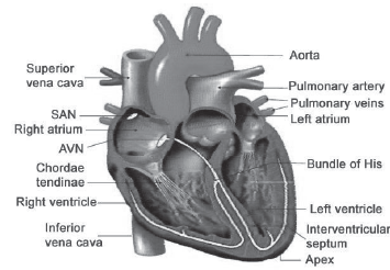
3. HEART
♦ It is a mesodermally derived organ located in mediastinum.
♦ It has the size of a clenched fist.
♦ It is protected by double-layered pericardium.
♦ The pericardial space (between pericardial membranes) is filled with pericardial fluid. It reduces the friction between the heart walls and surrounding tissues.
♦ Heart has 4 chambers- two upper atria (auricles) and two lower ventricles.
♦ The walls (cardiac muscles) of the ventricles are much thicker than that of the atria.
♦ The atria are separated by an inter-atrial septum and the ventricles are separated by inter-ventricular septum.
♦ In between atrium and ventricle, there is a thick fibrous atrio-ventricular septum with an opening.
♦ A tricuspid valve (3 muscular flaps or cusps) guards the opening between right atrium & right ventricle. A bicuspid (mitral) valve guards the opening between left atrium and left ventricle. These valves allow the flow of blood only in one direction, i.e. from atria to ventricles.
♦ Right ventricle has an opening to pulmonary artery and left ventricle has an opening to aorta. These openings have semi-lunar valves. They prevent backward flow of blood.
CONDUCTING SYSTEM OF HEART
♦ It includes nodal tissues, bundles & fibres.
♦ Nodal tissues are specialized cardiac musculature present in heart wall.
They are 2 types:
a. Sino-atrial node (SAN) in the right upper corner of the right atrium.
b.Atrio-ventricular node (AVN) in the lower left corner of the right atrium close to the atrio-ventricular septum.
♦ From the AVN, a bundle of fibrous atrio-ventricular bundle (AV bundle) passes through atrio-ventricular septa and divides into right & left branches. Each branch passes through the ventricular walls of its side. In the ventricular wall, it breaks up into minute fibres (Purkinje fibres). These fibres along with the bundles are known as bundle of His.
♦ Nodal tissues generate action potential without any external stimuli, i.e. it is autoexcitable. SAN initiates and maintains contraction of heart by generating action potentials (70-75/min). So, it is called the pacemaker.
CARDIAC CYCLE
It is the cyclic contraction and relaxation of heart for pumping blood. It involves 3 stages:
1. Joint diastole: It is the relaxed state of all chambers of heart. When the tricuspid and bicuspid valves open, blood from pulmonary vein and vena cava flows into left & right ventricles respectively through left and right atria. Semilunar valves are closed at this stage.
2. Atrial (Auricular) systole: SAN generates an action potential. As a result, both the atria contract. It is called atrial systole. This increases the flow of blood into the ventricles by about 30%.
3. Ventricular systole: The action potential is conducted to ventricular side by AVN & AV bundle from where bundle of His transmits it through the ventricular musculature. As a result, ventricles contract. It is called ventricular systole. During this, the atria undergo diastole. Ventricular systole increases the ventricular pressure causing
a. Closure of tricuspid and bicuspid valves due to attempted backflow of blood into the atria.
b. Semilunar valves open. So deoxygenated blood enters the pulmonary artery from right ventricle and oxygenated blood enters the aorta from left ventricle.
The ventricles now relax (ventricular diastole) and the ventricular pressure falls causing
a. The closure of the semilunar valves which prevents the backflow of blood into the ventricles.
b. The tricuspid and bicuspid valves are opened by the pressure in the atria.
The ventricles and atria again undergo joint diastole and the above processes are repeated.
A cardiac cycle is completed in 0.8 seconds.
♦ One heartbeat = a cardiac cycle.
So, normal heartbeat: 70-75 times/min (average: 72/min).
♦ Stroke volume: It is the volume of blood pumped out by each ventricle during a cardiac cycle. It is about 70 ml.
♦ Cardiac output: It is the volume of blood pumped out by each ventricle per minute, i.e. stroke volume x heart rate (70 x 72). It is about 5000 ml (5 litres). Cardiac output of an athlete is very high.
♦ Heart sounds: During each cardiac cycle, 2 sounds are produced. The first sound (lub) is due to the closure of tricuspid and bicuspid valves. The second sound (dub) is due to the closure of the semilunar valves.
One heartbeat = a lub + a dub.
ELECTROCARDIOGRAPH (ECG)
♦ It is an instrument used to obtain electrocardiogram.
♦ Electrocardiogram is the graphical representation of the electrical activity of the heart during a cardiac cycle.
♦ To get an ECG, a patient is connected to the machine with 3 electrical leads (one to each wrist and to left ankle) that monitor heart activity. For a detailed evaluation of heart’s function, multiple leads are attached to the chest region.
♦ An ECG consists of the following waves:
P-wave: Represents the excitation (depolarization) of atria during atrial systole.
QRS-complex: Represents depolarization of ventricles during Ventricular systole.
T-wave: Represents the repolarisation of ventricles.

Deviation in the ECG indicates the abnormality or disease. So, ECG has great clinical significance.
DOUBLE CIRCULATION
It is the circulation in which blood flows through the heart twice for completing its circuit. It includes:
1. Pulmonary circulation: Circulation b/w lungs and heart.
Deoxygenated blood from right ventricle → to pulmonary artery → to lungs → oxygenated blood → to pulmonary veins → left atrium.
2. Systemic circulation: Circulation b/w heart and various body parts.
Oxygenated blood from left ventricle → to aorta → arteries → arterioles → capillaries → tissues → deoxygenated blood from tissues → venules → veins → vena cava → to right atrium.
Systemic circulation provides nutrients, O2 and other substances to the tissues and takes CO2 and other harmful substances away for elimination.
♦ Hepatic portal system: It is a system which includes the hepatic portal vein that carries blood from intestine to the liver before it is delivered to the systemic circulation.
♦ Coronary circulatory system: It is a system of coronary vessels that circulate blood to and from cardiac musculature.

REGULATION OF CARDIAC ACTIVITY
♦ Normal activities of heart are auto-regulated by nodal tissues. So, it is called myogenic heart.
♦ Medulla oblongata regulates cardiac activity through ANS.
♦ Sympathetic nerves of ANS increase the rate of heartbeat, the strength of ventricular contraction and cardiac output.
♦ Parasympathetic nerves of ANS decrease the heartbeat, conduction of action potential and the cardiac output.
♦ Adrenal medullary hormones increase the cardiac output.
LYMPHATIC SYSTEM
♦ Includes Lymph, Lymph vessels & Lymph nodes (glands).
♦ As the blood passes through the capillaries in tissues, some water and soluble substances are filtered out from plasma to the intercellular spaces, to form tissue (interstitial) fluid. It has same mineral distribution as that in plasma.
♦ Some tissue fluid enters lymphatic system and the tissue fluid in them is called lymph. It drains back to major veins.
♦ Lymph is a colourless fluid containing lymphocytes.
Functions of lymph
♦ It is the middleman between blood & tissues. Tissue fluid helps to exchange nutrients, gases, etc. b/w blood and cells.
♦ It carries plasma proteins synthesized in liver to the blood.
♦ Transports digested fats (through lacteals in the intestinal villi), fat soluble vitamins, hormones etc.
♦ Filtration of bacteria and foreign particles.
♦ Lymph nodes produce WBC (lymphocytes) & antibodies.
DISORDERS OF CIRCULATORY SYSTEM
1. Hypertension (High Blood Pressure): The pressure of circulating blood on the walls of blood vessels is called blood pressure. Normal BP is 120/80 mm Hg. It includes systolic (pumping) pressure (120 mm Hg) and diastolic (resting) pressure (80 mm Hg).
When the BP is higher than normal, it is called hypertension. If an individual repeatedly has the BP of 140/90 or above, it shows hypertension. It leads to heart diseases and affects vital organs (brain, kidney etc).
2. Coronary Artery Disease (CAD) or Atherosclerosis: Here, Ca, fat, cholesterol and fibrous tissue are deposited in coronary arteries. So the lumen of arteries becomes narrower and thereby affects the blood supply.
3. Angina (angina pectoris): An acute chest pain due to O2 deficiency to heart muscles. It occurs due to improper blood flow. It is common among middle-aged and elderly.
4. Heart Failure (congestive heart failure): It is the inability of heart to pump blood enough to meet the needs of the body. Congestion of the lungs is the main symptom.
5. Cardiac arrest: Heart stops beating.
6. Heart attack: Sudden damage of heart muscle due to inadequate blood supply.